

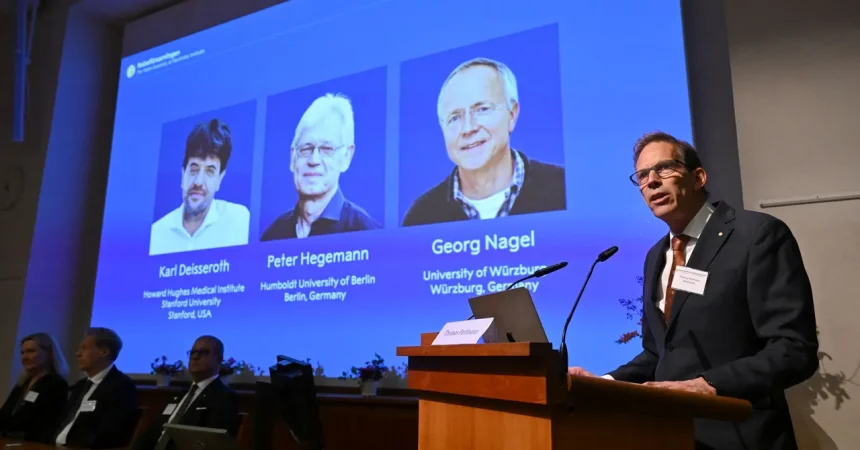
The 2026 Nobel Prize in Physiology or Medicine has been awarded to Karl Deisseroth of Stanford University, Peter Hegemann of Humboldt University in Berlin, and Georg Nagel of the University of Würzburg. The Karolinska Institute made the announcement, and the scientific community responded with a sense of wonder and gratitude. These three researchers are the architects of optogenetics, a technique that has given scientists something they long thought impossible: the ability to switch individual nerve cells on or off using beams of light. Per Svenningsson, chair of the Nobel Committee for Medicine, said, “Optogenetics provides opportunities for mapping the brain in a way that we could once only dream of.” That dream is now a daily reality in laboratories around the world. To understand why this matters, imagine trying to understand a symphony by listening only to the whole orchestra. You might notice patterns, but you could not tell which instrument plays which part, nor what would happen if one instrument fell silent. The brain is the most complex orchestra in existence, with billions of neurons firing in intricate rhythms. Optogenetics is like a conductor’s baton that can point to a single musician and ask them to play, or stop, at a precise moment. It lets researchers observe the living brain with extraordinary precision, revealing how circuits give rise to thoughts, emotions, and behaviors. It also helps them understand what happens when those circuits malfunction, offering new hope for treating neurological and psychiatric disorders. But the story behind this prize is not only about neuroscience. It is a powerful reminder that great discoveries often come from unexpected places, and that the boundaries between scientific disciplines are far more fluid than they seem. The roots of optogenetics lie in microbiology, in the study of a single-celled organism’s relationship with light. The path from pond scum to Nobel Prize is not a straight line, but it is a beautiful one.
Optogenetics has opened a window into the living brain that no other technique can match. Before this method existed, neuroscientists faced a frustrating problem. They could record the electrical activity of neurons, and they could observe which areas of the brain became active during different tasks, but they could not easily test cause and effect. If a certain group of cells fires during anxiety, is that firing actually causing the anxiety? To answer such questions, researchers needed a way to control specific cells, quickly and reversibly, without damaging the surrounding tissue. Optogenetics provides exactly that. By introducing light-sensitive proteins into selected neurons, scientists can make those cells respond to flashes of light. A pulse of blue light might make them fire; a pulse of yellow light might silence them. This is not a crude stimulation of a whole brain region. It is a precise, targeted intervention that can be delivered in milliseconds and turned off just as quickly. With this tool, researchers have begun to map the brain’s circuits in ways that were once unimaginable. They can trace the pathways involved in memory, motivation, sleep, pain, and social behavior. They can watch how the brain processes reward and punishment, and how it learns from experience. They can even investigate what goes wrong in conditions such as Parkinson’s disease, depression, addiction, epilepsy, and stroke. In animal models, optogenetics has already been used to restore movement in paralyzed limbs, to reduce compulsive behaviors, and to reactivate memories that seemed lost. The technique is not yet ready for widespread use in human patients, but it is an invaluable research tool, and it is already shaping the next generation of therapies. Perhaps most importantly, optogenetics embodies the power of convergence. It was born when microbiology, genetics, biophysics, and neuroscience came together. The discoveries that made it possible did not begin in the brain at all. They began in a tiny alga that had to find light to survive.
At the end of the last century, Peter Hegemann became fascinated by a single-celled green alga called Chlamydomonas. Chlamydomonas is not a plant, not an animal, but something in between. It has two flagella, tiny whip-like tails, that beat to move it through water. It is easy to overlook, but it is a master of survival. This humble organism lives in soil and freshwater, and it has a remarkable ability: it can sense light and move toward it. This is not a simple trick. For a single cell, finding the right light is a matter of survival, because light is the fuel for photosynthesis. Chlamydomonas has a tiny structure called an eye spot, a small orange dot on its surface. The color comes from retinal, a light-sensing molecule that is also found in the human eye. But the alga’s eye spot is far more primitive than ours. There is no lens, no retina, no complex wiring. It is just a patch of membrane packed with light-sensitive proteins. Hegemann wanted to understand how this simple structure could detect light so quickly and reliably. He used tiny electrodes to measure the electrical signals generated by the alga when it was exposed to light. What he found was astonishing. The alga produced an electrical impulse about half a millisecond after receiving light. The human eye, by comparison, takes at least ten milliseconds to begin its response. The alga was twenty times faster. This speed made no sense if the alga used the same elaborate chemical cascade that human eyes use. In our eyes, light hits a receptor, which triggers a series of biochemical steps, which eventually close or open ion channels. That process takes time. The alga was doing something much simpler and much more direct. Hegemann began to suspect that the eye spot contained a single protein that did everything: it absorbed the light, and it responded by opening a channel that let ions flow into the cell. This was a radical idea, and not everyone believed it.
In the early 1990s, Hegemann proposed that the eye spot of Chlamydomonas contained a protein that was both a light sensor and an ion channel. In other words, the same molecule detected the light and then acted on it, opening a pore in the cell membrane and allowing charged particles to rush through. This was a beautiful hypothesis, but it was also a controversial one. Science thrives on such skepticism. Extraordinary claims demand extraordinary evidence, and Hegemann had not yet provided it. He needed to find the molecule and prove its identity. Scientists had spent decades studying ion channels, and none of them could respond to light on their own. Ion channels are usually controlled by chemical signals, by changes in voltage, or by mechanical forces. The idea of a light-gated channel seemed almost too simple to be true. Hegemann knew that he needed evidence. He tried to isolate the light-sensitive proteins from the eye spot, hoping to study them in a test tube. But when the proteins were removed from their natural environment, they became unstable. They folded, clumped, or lost their function. It was like trying to understand how a fish swims by taking it out of water. The protein needed its home in the cell membrane, surrounded by the right lipids and proteins, to do its job. Hegemann’s team was stuck. Then a crucial breakthrough came from an unexpected place. A group of Japanese researchers sequenced the complete DNA of Chlamydomonas. This genetic blueprint provided a treasure map. Hegemann and his colleagues could now search for genes that might encode proteins with the expected characteristics of a light-sensing channel. They identified two genes that looked promising. These genes, they suspected, would produce proteins that could bind retinal and form a channel in the membrane. But a gene is just a set of instructions. To prove that these genes really did what they seemed to do, someone had to test them in a living cell. That is exactly what Georg Nagel did.
Georg Nagel took the next step, and it was a masterclass in experimental elegance. He took copies of the two genes identified by Hegemann’s team and inserted them separately into frog eggs. Frog eggs, particularly the eggs of the African clawed frog, are a classic tool in biology. They are large, easy to inject, and remarkably good at producing proteins from foreign genes. The eggs began to follow the instructions in the DNA, manufacturing the corresponding proteins and placing them in their outer membranes. Then Nagel did something wonderfully simple. He turned on the lights. When light hit the eggs, something extraordinary happened. The proteins in the membranes opened channels, allowing ions to flow across the membrane and generating an electrical current. The eggs had become light-sensitive. This was the proof that Hegemann’s hypothesis had been right. The experiment was so clean that it convinced even the skeptics. It showed that a single gene could confer light sensitivity on a cell that had never seen light before. This was not just a proof of concept; it was the birth of a new field. A single protein, now known as a channelrhodopsin, was enough to turn light into electrical activity. The discovery was a landmark in basic biology, but its full potential was not yet clear. It was Karl Deisseroth, a psychiatrist and bioengineer at Stanford, who saw the implications for neuroscience. If these light-sensitive proteins could be introduced into specific neurons in the brain, then those neurons could be controlled with light. Deisseroth and his colleagues took the channelrhodopsin genes and delivered them to neurons in living animals. Then they used thin optical fibers to shine light deep into the brain. When the light reached the neurons, the cells fired. When the light was turned off, they stopped. The technique was fast, precise, and reversible. It was optogenetics. Suddenly, scientists had a tool that could turn individual brain cells on and off with the flick of a switch.
The 2026 Nobel Prize in Medicine is a celebration of this achievement, but it is also a celebration of curiosity and collaboration. The prize also reminds us that the most useful tools are often born from the most basic questions. No one could have predicted that a pond alga would one day help us understand the human mind. But that is how science works: curiosity leads, and applications follow. Peter Hegemann did not set out to cure brain diseases. He was simply trying to understand how a tiny alga senses light. Georg Nagel did not set out to build a tool for neuroscience. He was testing a hypothesis about a protein. Karl Deisseroth did not set out to win a Nobel Prize. He was looking for better ways to understand the brain and to help people suffering from mental illness. Together, their work shows how fundamental research can lead to transformative applications. It also shows the importance of sharing knowledge across disciplines. The story begins with microbiology, moves through genetics and biophysics, and ends in neuroscience. Each step depended on the work of others, including the Japanese researchers who sequenced the alga’s genome. Science is not a solitary pursuit; it is a conversation that spans generations and continents. Today, optogenetics is used in laboratories around the world to study the brain and to develop new treatments for diseases. It has already deepened our understanding of Parkinson’s disease, depression, addiction, chronic pain, blindness, and spinal cord injury. It has allowed researchers to see how memories are formed and how they can be altered. It has given us a new language for talking about the mind. And it all began with a single-celled organism with an orange dot on its surface, a tiny eye spot that held a secret. The next time you see a patch of green pond water, you might remember that it contains more than algae. It contains a lesson about the power of patient observation, the courage to question accepted wisdom, and the astonishing connections that link all forms of life. Thanks to Hegemann, Nagel, and Deisseroth, we can finally switch on the lights in the darkest corners of the brain.